


 Copy to clipboard
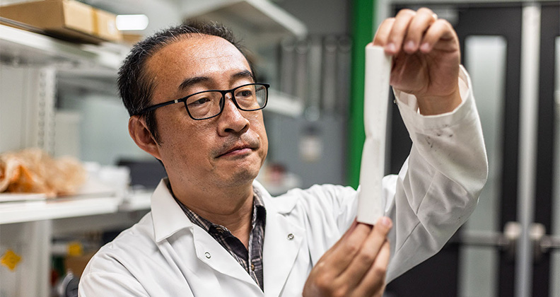
Mechanical engineers at The University of Texas at Dallas (UTD) collaborated with orthopedic surgeons at UT Southwestern Medical Center to develop a 3D-printed femur that’s designed to better prepare surgeons for complex procedures.
Wei Li, Ph.D., an Assistant Professor of Mechanical Engineering at UTD’s Erik Jonsson School of Engineering and Computer Science, led a team of researchers that set out to create a femur midsection that could be printed quickly and inexpensively. They published their first study on the 3D-printed femur in the August 2024 edition of the Journal of Orthopaedic Research.
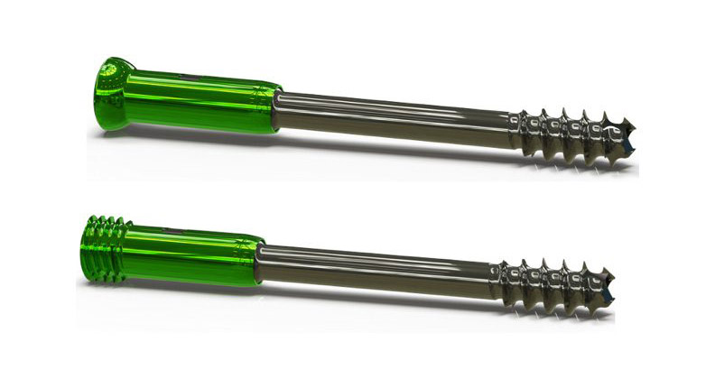
The current model of the bone mimics the dimensions and curvature of a human femur. Measuring approximately eight inches in length and one inch in diameter, the printed femur is made of polylactic acid (PLA), a biodegradable polymer frequently used in additive manufacturing (AM). Most notable is the low production cost: Each model can be printed for around $7.
According to Dr. Li, PLA could serve as a substitute for materials like titanium that are commonly used in bone repair. He said researchers might be able to print tumors onto the 3D-printed bones to test treatment options or use the models to support the growth of human bone tissue.
Dr. Li, who is also collaborating with NASA on 3D-printing technologies to replicate the surface of the moon, brings a unique engineering mindset to biomedical challenges.
“I’m interested in using an experimental approach to study 3D printing and using the multiple fixed models to simulate fundamental details of the printing and manufacturing processes,” he said.
Fine-tuning the Design
After countless iterations and nearly two years of trial and error, Dr. Li’s experimental AM process helped him and UTD mechanical engineering doctoral student Kishore Mysore Nagaraja print the femur model with the same geometry as real bone. The innovation holds promise for improving surgical outcomes; surgeons can use the printed bones to plan and rehearse procedures, minimizing uncertainty in the operating room.
Dr. Li’s team compared the 3D-printed model to real human donor bones, evaluating both for geometric accuracy and mechanical properties such as mature strength. By creating replicas of a patient’s unique anatomy, surgeons can better prepare for surgery and improve patient safety and surgical efficacy. With the 3D-printed bone as a guide, surgeons and researchers could apply various surgical plans with various tools to develop an optimal approach.
The process begins with preoperative imaging. “We use MRI or CT scans to capture a detailed 3D map of the patient’s bone structure,” Dr. Li explained. “From there, we print a prototype that precisely matches the patient’s anatomical geometry. Orthopedics is bioengineering this area of research, and 3D printing is a feasible technology for personalizing orthopedic implants.”
Dr. Li said that while the current application of this technology focuses on surgical training and educational modeling, he sees a broader and potentially more impactful application for its use: 3D printing sections of bone to replace and repair damaged bone in the human body. His team’s long-term goal is to combine biodegradable polymers like PLA with living femur cells or tissue to integrate the implant into the human body.
“AM opens the door to printing implants that can degrade safely,” he said. “Materials like PLA eventually break down into byproducts such as water and carbon dioxide.”
Step Toward Success
Despite its promise, 3D-printed bone replacement technology still faces significant obstacles before it can be used in clinical settings.
The first challenge is geometric accuracy. “We can acquire highly detailed 3D scans from MRI or CT, but there’s a limitation to the dimension of AM,” Dr. Li said. “To print the femur’s curved, complex surface, we need to use a very small printing diameter. Current manufacturing technology doesn’t allow for it.”
A second hurdle involves identifying suitable materials and optimizing the printing process to match the mechanical performance of real bone.
“We can use PLA to print bone structures, but so far there are no successful experiments that can prove that a PLA-printed structure can be used in the human body,” Dr. Li said. “We need more data to prove that this technology is safe and reliable for use in patients.”
One of Dr. Li’s most critical insights involves the need to integrate predictive modeling into the 3D printing process. By simulating the mechanical and structural outcomes before printing, engineers can better tailor each implant to clinical requirements.
The 3D-printed femur model is an example of how engineering innovation can transform clinical practice. From training surgeons to developing next-generation implants, Dr. Li’s work shows the potential of AM’s applications in orthopedics.
As AM continues to evolve, Dr. Li’s work represents a step toward improved surgical planning and the creation of regenerative bone implants. He noted that 3D printing is a complicated process with many printing parameters that need to be optimized.
He added, “Modeling that can help predict the geometric and mechanical properties of the printed bone structure needs to be built into the printing process to better meet the needs of hospitals, patients and the orthopedic market.”