In a recently published study, scientists working at the NYU Tandon School of Engineering and New York Stem Cell Foundation Research Institute announced that they’ve developed a way to produce artificial bone tissue that mimics the biological structures and complexities of naturally occurring bone tissue.
These replicas are precise enough to create an environment that allows for bone tissue growth from a person’s own mesenchymal stem cells, which could offer an exciting breakthrough for orthopedic implants and devices as well as drug discovery.
“Bone-mimetic surfaces, such as the one reproduced in this study, create unique possibilities for understanding cell biology and modeling bone diseases, and for developing more advanced drug screening platforms,” said Giuseppe Maria de Peppo, Ph.D., a Ralph Lauren Senior Principal Investigator at the New York Stem Cell Foundation Research Institute (NYSF) and the research team’s co-lead.
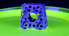
Researchers produced this artificial bone tissue using a nano-fabrication process called bio-thermal scanning probe lithography (bio-tSPL), which was developed in the lab run by team co-leader Elisa Riedo, Ph.D., Professor of Chemical and Biomolecular Engineering at the NYU Tandon School of Engineering.
This method involves using a “nano-chisel” and biothermal imaging on a biocompatible polymer to create a replica so detailed that it captures the minute, building-block components of actual bone tissue. The system allows for features in the bone tissue that are smaller than the size of a single protein or a billion times smaller than a meter. Using bio-tSPL is both cost-efficient and scalable, as these replicas can be reused multiple times.
“Until today, limitations in terms of throughput and biocompatibility of the materials have prevented [bio-tSPL’s] use in biological research,” Dr. Riedo said. “We are very excited to have broken these barriers and to have led tSPL into the realm of biomedical applications.”
Dr. Riedo told BONEZONE that one of the team’s immediate next steps involves focusing on understanding how stem cells sense the surroundings inside our body, and ultimately identify tissue environmental cues that improve differentiation of mesenchymal stem cells into mature osteoblasts, the cells that build our bones.
She added that the first step has been more focused on nano-fabrication, and on ways to scale up and decrease cost for the thermal scanning probe lithography method. “And the idea is, okay, now we know how to reproduce the bone; we know how to do it on the scale that [is] relevant for biological [and] for medical application.”
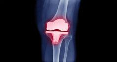
The timeline for clinical applications is still a work in progress. As Dr. de Peppo noted, any potential next steps are dependent upon what’s discovered in the coming years via research. However, the potential for safer and more effective bone therapies is strong. For example, he said, these cells could be used to treat people with fractures and bone defects that do not heal well. Also, we could transfer these bone environmental cues on orthopedic implants so that they integrate better with the bone tissue after implantation.
Annie Zaleski is a BONEZONE Contributor.


