


 Copy to clipboard
A new technology that produces 3D-printed bone structures containing living cells has been developed by scientists working at the University of New South Wales in Sydney, Australia. The technique, called ceramic omnidirectional bioprinting in cell suspensions (COBICS), could bring “unprecedented opportunities” in medicine, including the fabrication of autologous grafts using a patient’s cells and patient-specific, real-time bone reconstruction, according to Iman Roohani, Ph.D. He co-developed the technology with Associate Professor Kristopher Kilian, Ph.D.
“Overall, this technique may pave the way forward to how bone defects are managed after surgery, with scope for decreasing surgery time, greatly increasing bone healing time, and the potential to treat defects that have been impossible or at least challenging with earlier techniques,” said Dr. Roohani, whose background is in biomedical engineering, biomaterials and tissue engineering. “This approach may also eliminate the need for autologous grafts that are currently used for critical sized defects––that is, bone that has been lost due to injury or disease that will not regrow on its own.”
Current bone grafts are constructed with materials not suitable for living cells or are made with materials that don’t match native bone environments inside the body. The COBICS method builds structures with bone-like architecture and mechanics. The structures have the same chemistry of minerals found in bone, in the presence of biological molecules, without heating or toxic chemicals.
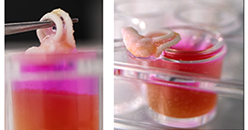
“Since the technique can be performed with living cells, we show that stem cells in the bath will immediately attach to the printed structure, proliferate, and begin to turn into bone cells just by virtue of the material,” Dr. Roohani said in an email. “In this way, our technique more closely emulates natural bone formation processes.”
The idea to create an ink in the form of a moldable paste that sets quickly in aqueous environments was inspired by the chemical and physical attributes of native bone minerals, said Dr. Roohani. “After ink optimization, we developed a special bath with what’s called yield-stress properties; that is, it behaves as a solid until an object shears into it at a certain force. We made a yield-stress material composed of microscopic hydrogel particles and living cells that you can print and convert into bone-mineral quickly after printing. The bath locks the ink where it is printed until it solidifies and becomes integrated with cells.”
The team at the University of New South Wales has begun testing the technology on animal subjects in the hopes the trials will produce clinical data needed for regulatory approval. Dr. Roohani cited challenges for bringing the technology to human surgical settings, including optimizing 3-D printers and other equipment. “We also would like to have enthusiastic orthopedic surgeons, dentists, or bone scientists to join and help us explore the potential of this technique for clinical applications,” he added.